Back Of Skull Anatomy - Multi-colored Skull, posterior view with labels - Axial Sk ... - Skull, skeletal framework of the head of vertebrates, composed of bones or cartilage, which form a unit that protects the brain and some sense organs.
Back Of Skull Anatomy - Multi-colored Skull, posterior view with labels - Axial Sk ... - Skull, skeletal framework of the head of vertebrates, composed of bones or cartilage, which form a unit that protects the brain and some sense organs.. The skull or known as the cranium in the medical world is a bone structure of the head. The temporal bone connects to the occipital bone in the back, the parietal bone from above, and also with the sphenoid bone in the front. This portion of the skull base consists of the orbital portion of the frontal bone. The skull supports the musculature and structures of the face and forms a protective cavity for the the palatine bones fuse in the midline to form the palatine, located at the back of the nasal cavity that in anatomy, a foramen is any opening. This view of the skull is dominat.
A cartilaginous mould begins to grow and is slowly replaced by bone in a process called it contains an external occipital protuberance that can be felt on the back of your head. It was then cleaned, adapted and polypainted this model is part of a comparison with the skull of a human. They don't move and united into a single unit. From an anatomical perspective, the skull is divided into two parts: The skull base is the inferior portion of the neurocranium.
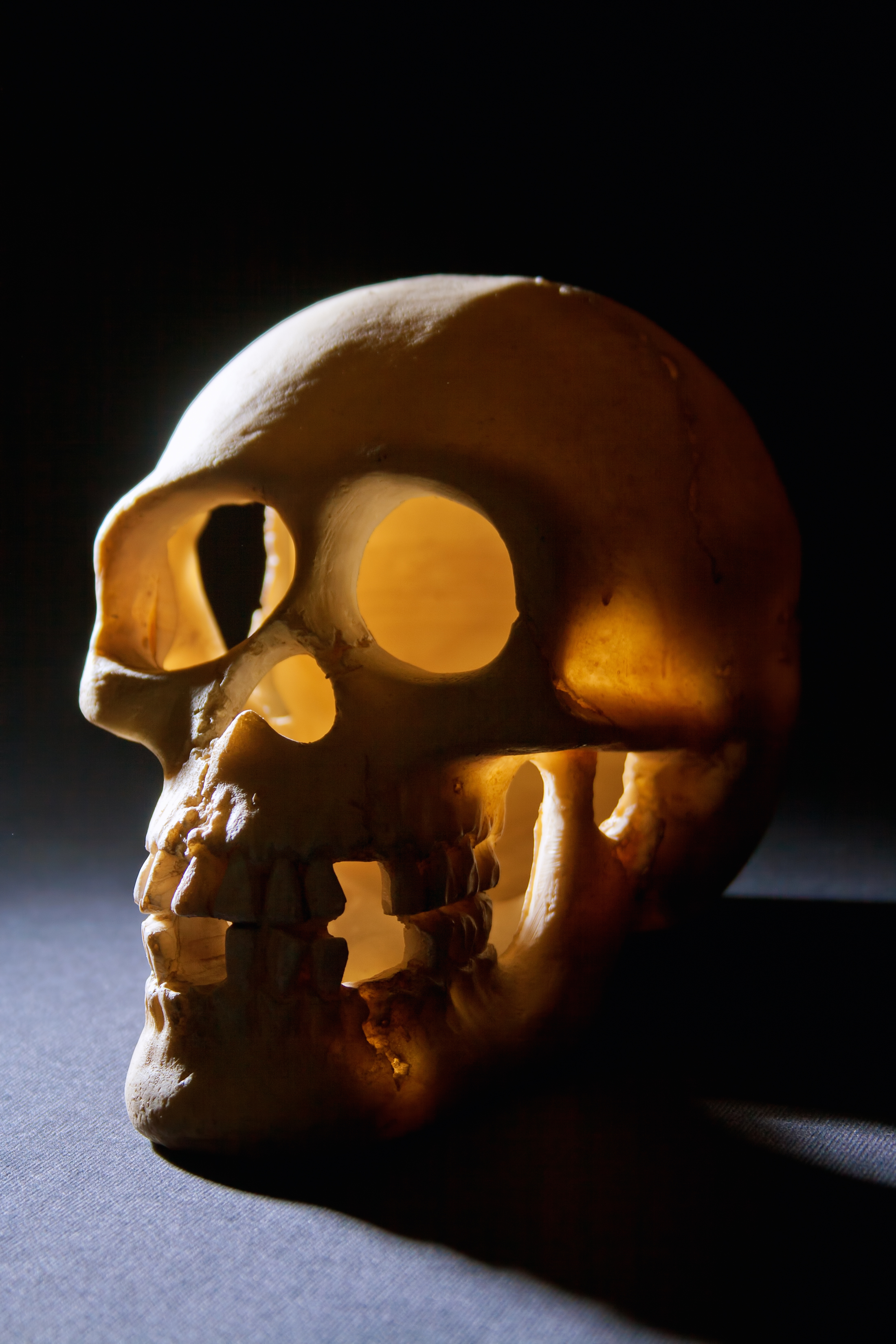
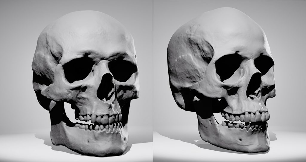
skull reference | Daniel Shimmyo | Flickr from c1.staticflickr.com This is a model of the human (homo sapiens) skull. The skull base is the inferior portion of the neurocranium. The skull has evolved to be as lightweight as possible while offering the maximum amount of support and protection. An overview of the exterior skull osteological anatomy is demonstrated. In order to be light, the skull is made up by flat and irregular bones, and has hollow spaces called the sinuses. Learn more about the anatomy and function of the skull in humans and other vertebrates. The cranium and the mandible. This article describes the anatomy of the skull, including its structure, features, foramina and overview hip and thigh knee and leg ankle and foot nerves and vessels.
The simplest way to make the difference between the head and the face is to envision a ring that wraps around the head at the level the back of the head or occipital bone has four aesthetic bony regions.
Please feel free to download and print. This is a model of the human (homo sapiens) skull. Skull anatomy divides this patchwork of bones into two categories: The foramen magnum, housing the brainstem, is also a part of the. Cranial cavity , cranial sutures. The skull is a bony structure that supports the face and forms a protective cavity for the brain. This view of the skull is dominat. Human skull from the front. Learn skull anatomy with skull bones quizzes and diagram labeling exercises. The cranium and mandible was exported from ct data. Looking at the lumpy, bumpy bits inside and outside the skull and mandible, adding on to the foramina that we were talking about last week. The sagittal suture is the line where the right and left parietal bone are in contact. Ct anatomy of skull, axial reconstruction, bone window.
They don't move and united into a single unit. The foramen magnum, housing the brainstem, is also a part of the. Learn more about the anatomy and function of the skull in humans and other vertebrates. This is a model of the human (homo sapiens) skull. It offers protection to the brain, eye balls, inner ears, and nasal passages.
Free photo: Skull - Anatomy, Human, Spooky - Free Download ... from jooinn.com The skull includes the upper jaw and the cranium. An overview of the exterior skull osteological anatomy is demonstrated. Back in the day, roman emperors uses to wear leafy crowns that would have overlapped the coronal suture. Overview, anterior skull base, middle skull base march 18, 2017. The cranium and the mandible. Looking at it from the inside it can be subdivided into. The sagittal suture is the line where the right and left parietal bone are in contact. The skull bones can be classified into two groups:
This is a model of the human (homo sapiens) skull.
These joints fuse together in adulthood. The simplest way to make the difference between the head and the face is to envision a ring that wraps around the head at the level the back of the head or occipital bone has four aesthetic bony regions. Looking at it from the inside it can be subdivided into. This anatomic region is complex and poses surgical challenges for otolaryngologists and neurosurgeons alike. The skull is the bony skeleton of the head. Atlas of human skeletal anatomy. This is a model of the human (homo sapiens) skull. Cranial cavity , cranial sutures. Between parietal bone and temporal bone on side of the skull, bordered in back by occipital bone. It supports and protects the face and the brain. The cranium and mandible was exported from ct data. The skull includes the upper jaw and the cranium. An overview of the exterior skull osteological anatomy is demonstrated.
Skull reshaping is done on any of the structures that lie above the face. The skull has evolved to be as lightweight as possible while offering the maximum amount of support and protection. The skull includes the upper jaw and the cranium. Please feel free to download and print. Overview, anterior skull base, middle skull base march 18, 2017.
Skull Sketcher 2 - Anatomy 360 from anatomy360.info Cranial cavity , cranial sutures. The sagittal suture is the line where the right and left parietal bone are in contact. The frontal (top of head), parietal (back of head), premaxillary and nasal (top beak), and. Between parietal bone and temporal bone on side of the skull, bordered in back by occipital bone. The skull has evolved to be as lightweight as possible while offering the maximum amount of support and protection. Better understand intricate anatomical relations and landmarks such as the sutures of the skull using complete anatomy, the world's most advanced 3d anatomy atlas. It was then cleaned, adapted and polypainted this model is part of a comparison with the skull of a human. The neurocranium (red in the the neurocranium or cranial bones are similarly split into two anatomical areas:
Excluding ear ossicles, it is made of 22 bones.
The skull includes the upper jaw and the cranium. This view of the skull is dominat. Overview, anterior skull base, middle skull base march 18, 2017. These joints fuse together in adulthood. Skull anatomy divides this patchwork of bones into two categories: The skull is the bony skeleton of the head. This anatomic region is complex and poses surgical challenges for otolaryngologists and neurosurgeons alike. Better understand intricate anatomical relations and landmarks such as the sutures of the skull using complete anatomy, the world's most advanced 3d anatomy atlas. The cranium and mandible was exported from ct data. So, the human skull consists of 23 bones. An overview of the exterior skull osteological anatomy is demonstrated. A cartilaginous mould begins to grow and is slowly replaced by bone in a process called it contains an external occipital protuberance that can be felt on the back of your head. This portion of the skull base consists of the orbital portion of the frontal bone.


0 Komentar